Reproduced from Shutterstock by Subscription
It was a Monday and I was the intern at Tufts who had been on Sunday. That meant I had gone in Sunday morning and worked Sunday night and all day Monday until my work was done. We were generally on every third night except when we were on the famous Louis Weinstein’s infectious disease service, which was every other night for two months a year. We also had one month at the since-closed Boston Veterans Administration Hospital which was every fourth night. That was so easy we called our month there “time at the VA spa”. I am telling you this because I want you to know that I really wanted to go home that Monday evening.
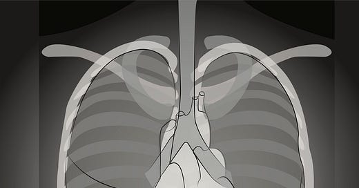
During the day Monday I had admitted a woman who had had over 10 admissions for pancreatitis. She came in again with her usual abdominal pain so I assumed she had “gotten it again”. 1 I ordered the usual tests including a chest X-ray which was routine for most admissions. I proceeded to attend to my other patients and tasks. I was ready to leave at about 6:30 pm when I realized I had not seen her chest X-ray. Radiology read the X-rays, but did not often call us for abnormalities, if ever. We were expected to review what we ordered. This was also 1973, so way before you could pull up the image on a computer. I wanted to leave, but schlepped down to Radiology and pulled up my patient’s film. They were located on a revolving film holder which you rotated until you reached your patient. When my patient’s film came up, I thought there was air under the right diaphragm. I verified this with the radiology resident and called surgery. She was operated on emergently and was found to have perforated her duodenum. I looked up “pancreatitis and duodenal perforation” in preparing this blog. There are only rare reports of this combination.
Wow, did I ever learn the value of an upright chest X-ray in abdominal pain, and that experience ultimately lead to one of my rules: Try always to get the chest X-ray with the patient in an upright or semi-upright position.
Supine chest X-rays are useful in critically ill patients to determine the location of central lines and endotracheal tubes, but even then as mentioned below, upright is better. Supine films are better than nothing if the patient absolutely cannot sit up. At all other times, avoid getting supine chest X-rays because they provide much less information than an upright film. Here’s what you can miss with a supine chest X-ray:
1. Vascular redistribution since the chance to observe engorgement of the lung field’s upper vessels is lost. Upper vessel vascular congestion is classic for heart failure.
2. Pleural effusions since the fluid layers out evenly in a flat chest X-ray
3. A pneumothorax since with a supine film, the free air rises to the anterior chest where it layers out and is difficult to detect. In an upright film the free air rises to the lungs’ apices where it creates a contrast with the lung below. Detecting a pneumothorax is especially important after placement of a central line or pacemaker and in intubated patients. These patients are at increased risk for a pneumothorax from the barotrauma associated with mechanical respiration and increased inhalation pressures.
4. Free air under the diaphragm from perforation of an abdominal viscus.
For all of these reasons, flat chest X-rays should be avoided in general and in critically ill patients in particular. There is much more information in an upright film. So check whether the X-ray was flat or upright before you conclude that there is no vascular redistribution or pleural effusion in your heart failure patient or that there is not pneumothorax after an internal jugular or subclavian vein cannulation.
That Isaac Newton guy was probably onto something!
1. https://pauldthompsonmd.substack.com/p/why-you-too-should-be-cheap?utm_source=profile&utm_medium=reader2
#chest X-ray #pneumothorax #pleural effusion #heart failure #free air
#pneumothorax #pleural effusion #heart failure #chest X-ray


